
Royal Dental College
E-ISSN: Coming Soon
E-ISSN: Coming Soon
Surgical Extraction of Mesiodens under general Anesthesia- A Case Report
Full Html
INTRODUCTION
Supernumerary teeth are additional teeth that develop beyond the normal dentition and can be found in both the primary and permanent dentitions, with a slight predilection for occurrence in the anterior region of the maxilla.[1] The prevalence of supernumerary teeth (SNT) in general population is estimated to be 0.1–1%, with no significant sex- or age-related predilection.[2] The majority of supernumerary teeth are associated with the permanent dentition and are most commonly located in the pre maxillary midline region, where they are referred to as mesiodens, of these, most are reported to remain unerupted.[3,4] This case highlights the management of an inverted, conical-shaped rudimentary supernumerary tooth (mesiodens) located in the midline of the premaxilla in an 8-year-old male patient. The case is notable for its proximity to the nasal cavity and the use of a labial surgical approach, which is relatively uncommon compared to the traditional palatal or intraoral techniques. The report discusses the clinical presentation, radiographic findings, and surgical decision-making process, emphasizing the advantages of the chosen approach in this specific anatomical and developmental context.
CASE REPORT
An 8-year-old male child reported to the Department of Pediatric and Preventive Dentistry, Royal Dental College, Chalissery with a chief complaint of additional tooth in upper front tooth region since 1 year. He was a healthy child with a non-contributory medical and dental history. An initial examination conducted one year earlier revealed an asymptomatic maxillary midline supernumerary tooth visible in the oral cavity and an inverted maxillary mesiodens impacted in the midline near the nasal cavity. Clinical examination also showed impacted left and right permanent lateral incisors. (Fig 1)
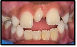
Fig 1: Preoperative photograph showing mesiodens in between tooth 11 and 21
.png)
Fig 2: Anterior view of CBCT
.png)
Fig 3: Posterior view of CBCT
For surgical evaluation and to determine the exact location and orientation of the tooth, the patient underwent cone-beam computed tomography (CBCT) imaging. CBCT revealed an vertically erupted supernumerary tooth between 11 and 21 and inversely and vertically impacted supernumerary tooth with dilaceration and enlarged follicular space. (Fig 2 & 3) Extraction was planned for the mesiodens with regular follow up till the laterals erupts.
.png)
Fig 4: CBCT revealing the distance of mesiodens from the anatomical landmarks
The tooth was confirmed to be palatally positioned. (Fig 4) With parental consent, the decision was made to proceed with surgical removal of the mesiodens under general anaesthesia. A labial surgical approach was planned for the procedure as the proximity of the mesiodens to the nasopalatine canal, the buccal approach was selected to minimize the risk of nasopalatine nerve injury.
TREATMENT
The mesiodens erupted in between tooth 11 and 21 was extracted normally. (Fig 5) For extraction of impacted inverted mesiodens, a crestal incision was made using a #15 Bard-Parker blade extending from the distal aspect of the maxillary right lateral incisor to the distal aspect of the maxillary left lateral incisor. (Fig 6) A full-thickness mucoperiosteal flap was then elevated with a periosteal elevator. A slight alveolar prominence was noted between the apices of the central incisors, which, upon palpation, corresponded to the anterior extent of the mesiodens. Using a sterile #8 round bur at slow speed, the buccal cortical plate was carefully removed under copious saline irrigation to expose the tooth. A circular bony window was created around the mesiodens with caution to avoid excessive bone removal and to prevent injury to the adjacent roots. The tooth was found to be vertically oriented, with its root apex positioned labially and the crown directed superiorly. Partial sectioning of the mesiodens was performed to facilitate luxation; however, removal was initially hindered by the crown’s size. (Fig 7) After further circumferential enlargement of the bony window, the tooth was successfully delivered.
.png)
Fig 5: Extracted mesiodens in between permanent central incisors
The extraction site was gently curetted, irrigated with saline and betadine, and closed with 11 interrupted sutures. Postoperative pain was managed with analgesics and antibiotics. The patient’s recovery was uneventful, with no unusual signs or symptoms reported.
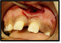
Fig 6: Crestal incision was made using a #15 Bard-Parker blade
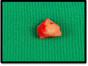
Fig 7: Extracted inverted impacted mesiodens
A follow-up examination conducted one week postoperatively revealed satisfactory healing, and suture removal was performed at three weeks, showing normal tissue repair at the surgical site. (Fig 8)
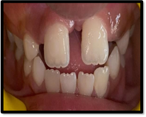
Fig 8: Postoperative review after 3 weeks
One month after extraction, the patient was recalled, and stainless-steel retainers were placed, attached to brackets and E-chain on the right and left central incisors, to facilitate space closure between the centrals and promote eruption of the lateral incisors. (Fig 9)
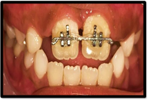
Fig 9- space closure initiated after placement of bracket and E- chain
Following bracket placement, the patient was reviewed weekly. Complete space closure was achieved within two weeks, after which lingual retainers were bonded to maintain the achieved tooth position. (Fig 10) The patient is currently under review and is planned for further orthodontic intervention to address tongue thrusting and mouth- breathing habit and to correct the anterior open bite.
.png)
Fig 10: Space closure achieved in between central incisor followed by placement of lingual retainer
DISCUSSION
Supernumerary teeth are developmental anomalies defined as any tooth or odontogenic structure that arises from tooth buds in excess of the usual number for a given region of the dental arch.[5] The presence of a mesiodens near the nasal cavity is a rare clinical finding. Supernumerary teeth are broadly classified into two types: supplemental (tooth-like) and rudimentary. The aetiology of supernumerary teeth remains unclear; however, the most widely accepted theory suggests hyperactivity of the dental lamina as the causative factor (Primosch, 1981). Although a hereditary component and a male predilection have been proposed (Brunning et al., 1957; Sedano and Gorlin, 1969), these associations have yet to be definitively established.[6] Studies conducted during the early mixed dentition stage indicate that although mesiodens are present, they fail to erupt in 79–91% of cases. In 7–20% of the cases analysed, their presence is identified incidentally on routine radiographs without any associated pathology. These findings confirm that the mixed dentition period is the stage when impacted mesiodens are most commonly diagnosed and underscore the importance of routine dental radiographic examinations.[5] The most frequent cause of impacted maxillary incisors is the presence of supernumerary teeth. Furthermore, evidence suggests that tuberculate and odontome types are more likely to obstruct the eruption of permanent maxillary incisors compared to other types. Due to the absence of clinical signs in most cases, impacted mesiodens are often detected incidentally on two-dimensional (2D) radiographs such as orthopantomographs (OPGs), occlusal, or periapical radiographs, and, in some instances, even lateral cephalometric radiographs. However, Cone-Beam Computed Tomography (CBCT) has emerged as a valuable diagnostic tool that significantly enhances the accuracy of detection by providing three-dimensional (3D) cross-sectional images.[7] The utilization of CBCT has recently gained prominence as a highly effective modality for the evaluation of mesiodens. It allows comprehensive and precise visualization of mesiodens and the surrounding anatomical structures, thereby enabling clinicians to select the most appropriate surgical approach and minimize trauma to adjacent teeth and vital structures. Additionally, a study by Maddalone et al. advocated the use of digital radiography, due to its low radiation exposure, for screening primary school children especially those with a family history of mesiodens even in the absence of clinical symptoms.[8] The authors emphasized that early detection and timely intervention are essential to prevent serious complications such as root resorption and displacement of adjacent teeth. Mesiodens have been associated with various syndromic conditions, including cleft lip and palate, cleidocranial dysostotic, Gardner’s syndrome, and chondroectodermal dysplasia.[9] They may also contribute to several dental complications, such as impaction or ectopic eruption of adjacent teeth, cyst formation, crowding, displacement, rotation of neighbouring teeth, and root resorption.[10] Furthermore, in patients with cleft palate, the presence of mesiodens can compromise the success of secondary alveolar bone grafting and subsequent implant placement.[11] General anaesthesia is often the preferred modality for extracting mesiodens in young or behaviourally uncooperative children, ensuring both safety and procedural efficiency.[12] Under General anaesthesia, children remain unconscious and immobile, allowing clinicians to perform extractions precisely while reducing the risk of psychological trauma. Typical management may include mask induction with sevoflurane and maintenance through inhalational agents or intravenous access, often supplemented by local infiltration to control intraoperative pain.[13] This approach not only establishes a controlled airway via endotracheal intubation or laryngeal mask airway, but also significantly decreases the likelihood of intra- and postoperative complications, making it a highly effective strategy in paediatric dental care.[14] The palatal approach is the most commonly reported technique for the extraction of impacted mesiodens in the literature, followed by the buccal approach.[15] The conventional buccal approach offers excellent surgical visibility; however, in cases of inverted mesiodens, it often necessitates extensive osteotomy, which can increase the risk of intraoperative complications and postoperative sequelae, such as damage to the roots of adjacent permanent incisors.[16] Additionally, excessive bone removal associated with this approach may lead to heightened postoperative swelling and pain. To circumvent these challenges, the palatal approach is often considered a viable alternative; however, it carries a significant risk of injury to the nasopalatine nerve, particularly when the mesiodens is positioned anterior to the nasopalatine canal.[6] In the present case, given the proximity of the mesiodens to the nasopalatine canal, the buccal approach was selected to minimize the risk of nasopalatine nerve injury. There are indications for both early surgical removal of supernumerary teeth and their supervised observation. The case presented involved a mesiodens that showed no change in position over a reasonable period of time. Its presence resulted in the formation of a diastema between the central incisors and altered the eruption path of the permanent lateral incisors. Given these factors, significant spontaneous improvement in the mesiodens position was unlikely, and surgical removal was deemed the most appropriate course of action.[5]
CONCLUSION
This case highlights the critical role of thorough clinical and radiographic evaluation, emphasizing interdisciplinary collaboration between medicine and dentistry to address patient needs effectively and deliver comprehensive care aimed at improving quality of life. Within the limitations of this report, the intraoral approach via the nasal floor for the surgical removal of inverted mesiodens proves to be a predictable, safe, and time-efficient technique. When applied appropriately, it minimizes the risk of intraoperative and postoperative complications and significantly reduces postoperative swelling and pain.
References
1. Kumar V, Bhaskar A, Kapoor R, Malik P. Conservative surgical management of a supernumerary tooth in the nasal cavity. BMJ Case Reports CP. 2020 Jul 28;13(7):e235718. doi: 10.1136/bcr-2020-235718.
2. Rahman S, Madan T, Goyal RJ, Goyal S, Bhalla V Prabhu MS. The Teeth in the Nasal Cavity. Bengal Journal of Otolaryngology and Head Neck Surgery.2024;32(2):99-103.
doi.org/10.47210/bjohns.2024.v32i2.100.
3. Nazif MM, Ruffalo RC, Zullo T. Impacted supernumerary teeth: a survey of 50 cases. J Am Dent Assoc. 1983 Feb;106(2):201-4. doi: 10.14219/jada.archive.1983.0390.
4. Tay F, Pang A, Yuen S. Unerupted maxillary anterior supernumerary teeth: report of 204 cases. ASDC Journal of Dentistry for Children. 1984 Jul 1;51(4):289-94.
5. Henry RJ, Post AC. A labially positioned mesiodens. Case report. Pediatric Dentistry. 1989 Mar;11(1):59-63.
6. Urechescu H, Banu A, Streian F, Urtila F, Cuzic C, Dinu S et al. Intraoral Approach Through the Nasal Floor for Surgical Removal of Inverted Mesiodens: Protocol and Case Series. J Clin Med. 2024 Dec 22;13(24):7831. doi: 10.3390/jcm13247831.
7. Mossaz J, Kloukos D, Pandis N, Suter VG, Katsaros C, Bornstein MM. Morphologic characteristics, location, and associated complications of maxillary and mandibular supernumerary teeth as evaluated using cone beam computed tomography. Eur. J. Orthod. 2014 Dec;36(6):708–718. doi: 10.1093/ejo/cjt101.
8. Maddalone M, Rota E, Amosso E, Porcaro G, Mirabelli L. Evaluation of surgical options for supernumerary teeth in the anterior maxilla. Int J Clin Pediatr Dent 2018; July-Aug;11(4):294-298. doi: 10.5005/jp-journals-10005-1529.
9. Aoun G, Nasseh I. Mesiodens within the nasopalatine canal: an exceptional entity. Clin Pract. 2016 Dec 7;6(4):903. doi: 10.4081/cp.2016.903.
10. Bin SE, Roby PM. An Inverted Impacted Mesiodens Perforating the Nasal Floor with an Impacted Canine. Int J Oral Dent Health. 2019;5(1):082. doi:10.23937/2469-5734/1510082.
11. Karim ZA, Musa N, Noor SN. Utilization of dental general anesthesia for children. Malays J Med Sci: 2008 Jul;15(3):31-9.
12. Campbell RL, Shetty NS, Shetty KS, Pope HL, Campbell JR. Pediatric dental surgery under general anesthesia: uncooperative children. Anesth prog. 2018 Jan 1;65(4):225-230. doi: 10.2344/anpr-65-03-04.
13. Puri S, Mathew PJ. General Anesthesia for Dental Procedures in Children: A Comprehensive Review. J Postgrad Med Edu Res. 2022 Feb 19;56(1):29-33. doi: 10.5005/jp-journals-10028-1555.
14. Kong J, Peng Z, Zhong T, Shu H, Wang J, Kuang Y et al. Clinical analysis of approach selection of extraction of maxillary embedded mesiodens in children. Dis Markers. 2022 May 3;2022(1):6517024. doi: 10.1155/2022/6517024.
15. Kim Y, Jeong T, Kim J, Shin J, Kim S. Effects of mesiodens on adjacent permanent teeth: a retrospective study in Korean children based on cone‐beam computed tomography. Int J Paediatr Dent. 2018 Mar;28(2):161-9. doi: 10.1111/ipd.12317.
16. Omami M, Chokri A, Hentati H, Selmi J. Cone-beam computed tomography exploration and surgical management of palatal, inverted, and impacted mesiodens. Contemp Clin Dent. 2015 Sep 1;6(Suppl 1):S289-93. doi: 10.4103/0976- 237X.166815.
